Spectator

joined 1 year ago
MODERATOR OF
 

Patient was a young adult working in finance at a major tech company found to be mute and diaphoretic.

Physical exam notable for fever, tachycardiac, hypertension, awake but not following commands, aphasic, and with hyperreflexia and muscle ridigity. CK peaked to 11,344.

MRI shows multiple ovoid to splotchy confluent lesions in the white matter with diffusion restriction. Lesions also enhanced with hyperperfusion (not shown).

Urine drug test positive for cocaine. Infectious work-up was negative. Steroids were started with good recovery.

Patient denied knowingly taking cocaine but did say weekly use of what they thought was MDMA with friends...

Final diagnosis: Levamisole-induced leukoencephalopathy. Levamisole is an antiparasite medication that is no longer used in the US but still in some other countries. It is a common cutting agent in cocaine. It's neurotoxic effects primarily come from causing demyelination.


I remember this episode quite well because it happened around the time I decided to get into the medical field. In the episode, a young teacher had a first-time seizure while in the middle of teaching. House and team attempted to get a brain MRI, but she got an allergic reaction from the IV contrast. Thereafter, some drama happens, and at some point, they break into her house, find out she's been eating raw pork (wtf?), and diagnose her with the tapeworm infection associated with eating raw pork, cysticercosis (and neurocysticercosis, since it also involved her brain). They took an x-ray of her leg to show all the parasites in the muscles, and then House scolds her for being stupid. I remember thinking that was such as crazy medical story.

The reality is - they could have just repeated the brain MRI minus the contrast part, and the radiologist would have been able to identify neurocysticercosis without issue. House would have complained to Cuddy that she really was wasting his time with these basic cases, and the episode would have lasted 15 minutes tops...

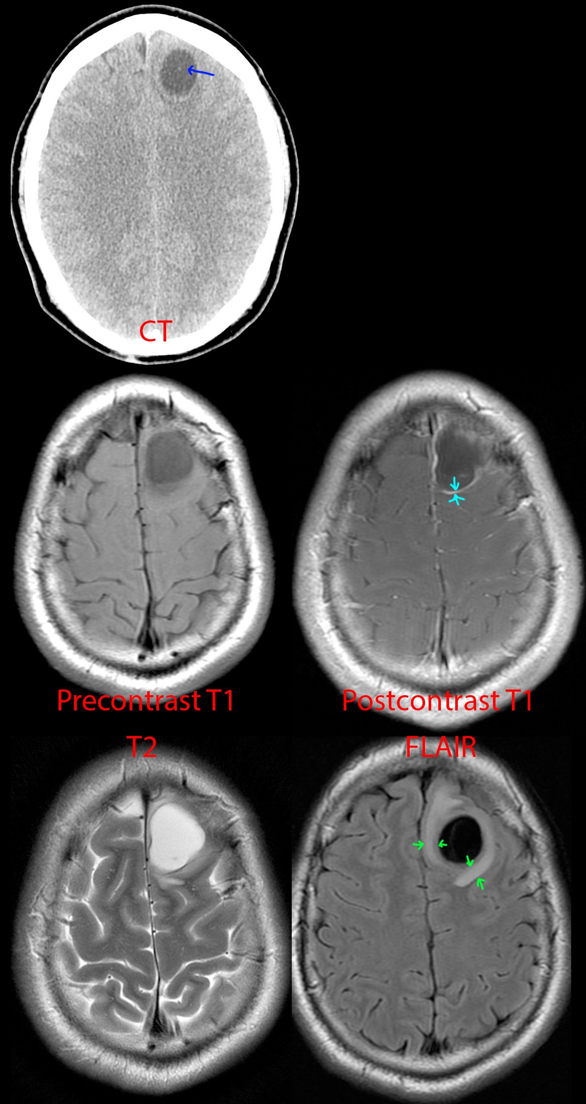
Anyhow, this is a 25 year old Hispanic from jail. Just like the House episode, he presented with first time seizure and headaches.

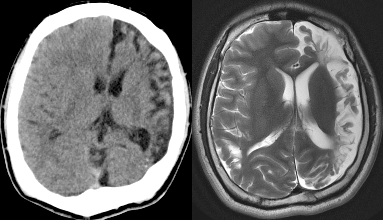
CT of the head [top] shows a cystic lesion in the left frontal lobe. If one pays attention, one can see a small dot (blue arrow) within the cyst representing the scolex of the tapeworm parasite. Just from the CT appearance, history of seizure, and risk factors of jail (the parasite thrives in areas of low sanitation) and Hispanic (the parasite is endemic to South America), neurocysticercosis is the top possibility. A differential diagnosis of cystic brain tumor is provided to complete the picture.

MR [middle and bottom] shows a cystic lesion again. After giving IV contrast [middle right], one can see the cyst has a thin wall of enhancement (teal arrows). On T2 [bottom left] and especially FLAIR [bottom right], one can see a rim of swollen brain (green arrows) from the inflammation going on around the parasite.

This was diagnosed as neurocysticercosis in the colloidal vesicular stage and antiparasite medication was started.


[Top]: X-ray shows a lucent, bubbly, lesion of the distal femur at the metaphysis. On the frontal view [top right], there is breakage through the medial femoral cortex into the adjacent soft tissues, not a good sign.

[Bottom]: MRI shows a multicystic lesion filling the distal femur containing multiple locules, many with fluid-fluid, fluid-debris, and fluid-hemorrhage levels. The most common lesions with this striking appearance are aneurysmal bone cyst, giant cell tumor, or telangiectatic osteosarcoma. Unfortunately, there is clearly extension of the bone tumor beyond the bone (yellow arrows), which favors a more aggressive neoplasm from that differential diagnosis - this turned out to be telangiectatic osteosarcoma.


5 year old who fell off a slide.

Initial imaging shows a comminuted fracture through the distal humerus, compatible with a supracondylar fracture. Nothing else appreciable here, except maybe in retrospect some lucency of the distal humerus where the fracture is.

4- and 7-month follow-up radiographs shows a growing lucent lesion of the distal humerus, expanding the bone there. It has a multicystic appearance. A diagnosis of large simple bone cyst versus aneurysmal bone cyst was proposed.

12 month follow-up was done after the cyst was opened surgically, its contents scraped off, and the resulting cavity was packed with allograft bone chips. At surgery, this turned out to be an aneurysmal bone cyst.

5 year follow-up shows involution of the cyst cavity with some residual heterogeneity and a bone spur at the anterior aspect of the distal humerus.


[Left]: Head CT shows left hemispheric volume loss. The injury happened early enough that even the skull is smaller on that side.

[Right]: Brain MRI shows the severe left hemispheric atrophy. Some of the brain gyri have bulbous ends and a thin neck, resembling mushrooms, a shape called ulegyria and consequence of the brain atrophy. The left lateral ventricle is mildly enlarged due to the atrophied brain.


Red arrows point to 2 big gallstones, top one in the gallbladder and bottom one obstructing a small bowel loop, and a small gallstone in the cystic duct.


Red lines point to hernia entry. Red arrow points to where the bowel tapers and becomes obstructed as it enters the hernia sac.


[Left]: Fetal MRI (FIESTA sequence) shows twins joined from their lower chest to the pelvis, but truly fused and sharing a single abnormal pelvic region. Not shown, but there are 3 lower limbs - one of the twins only had a single lower extremity.

[Right]: Postnatal small bowel follow-through (SBFT). It was unclear initially whether the twins shared a single rectum or had their own rectum. Therefore, contrast was administered via nasogastric tube for the twin with the suspected nonfunctional rectum, and serial imaging was performed until it passed into what turned out to be a separate, functional, but small rectum/anus.

I do not know too much about conjoined twins - not my area of expertise, but the general forms to consider are the side of fusion: ventral (front to front), lateral (side to side), dorsal (back to back), or caudal (tail end to tail end). Within these first 3, there are subtypes depending on how far up the fusion goes (head, chest, abdomen/pelvis); by definition, the caudal version obviously is only a lower body fusion. Once this is derived, an additional classification is the number of upper and lower limbs.


No clinical history saved on this one - sorry.

[Right] Small bowel follow-through (SBFT), where the patient drinks barium, and then we wait a bit until that barium is in the small bowel, then we take some pictures. This study is showing a long segment of terminal ileum that is strictured and severely narrowed in fibrostenotic Crohn's (red bracket). This is called the "string sign."

[Left] Coronal CT performed sometime after the SBFT. You can still see some residual barium in the small and large bowels (blue arrows). Red bracket shows the CT appearance of the terminal ileum stricture. On the CT, you can also see that the strictured segment has submucosal fat deposition, the "fat halo sign."


I didn't save any clinical history for these - sorry.

[Top] Patient 1 - Gigantic mass along the lesser curvature of the stomach. Look down at your belly - this mass is about 1/3rd the width from left to right.

[Mid] Patient 2 - CTs showing gently lobulated and undulating wall thickening of the gastric cardiac and fundus. Notice the transition from the normal gastric rugae to the smoother wall thickening where it is infiltrated by lymphoma. There is also mild (aneurysmal) dilation of the stomach where the wall thickening is located.

[Bottom] Patient 2 - PET-CT. The wall thickening is ridiculously hypermetabolic with a max SUV of 21.3. For comparison, the liver is normally in the range of 2-4 mean SUV.

Tuberculosis, sarcoidosis, lymphoma, and metastatic disease - these 4 can look like almost anything.

1
submitted 1 year ago* (last edited 1 year ago) by Spectator@lemmy.world to c/radiology@lemmy.world
 

57 year old with left flank pain for 12 hours. Urine sample 2+ for blood. White count at 14.

[Top]: Coronal CT shows the left kidney is enlarged, with angry-looking, inflamed, surrounding fat. The renal pelvis is dilated (hydronephrosis).

[Bottom]: Axial CT shows a small stone at the very end of the ureter (ureterovesicular junction).

Pretty straightforward case. For the nonmedical visitors, this is what we look for if your doc wants to get a CT for flank pain / kidney stone suspicion. See the other case for an ultrasound version.

1
submitted 1 year ago* (last edited 1 year ago) by Spectator@lemmy.world to c/radiology@lemmy.world
 

25 year old with right flank and pelvic pain for 2 days. No fevers, chills, nausea, vomiting, or other urinary symptoms however.

[Top]: Ultrasound shows right kidney with distended pelvis (hydronephrosis).

[Mid & Bottom]: Ultrasound transverse and sagittal images shows a 4.5 mm stone at the very end of the ureter (ureterovesicular junction).

Pretty straightforward case. For the nonmedical visitors, this is what we look for if your doc wants to get an US for flank pain / kidney stone suspicion. See the other case for a CT version.

view more: ‹ prev next ›