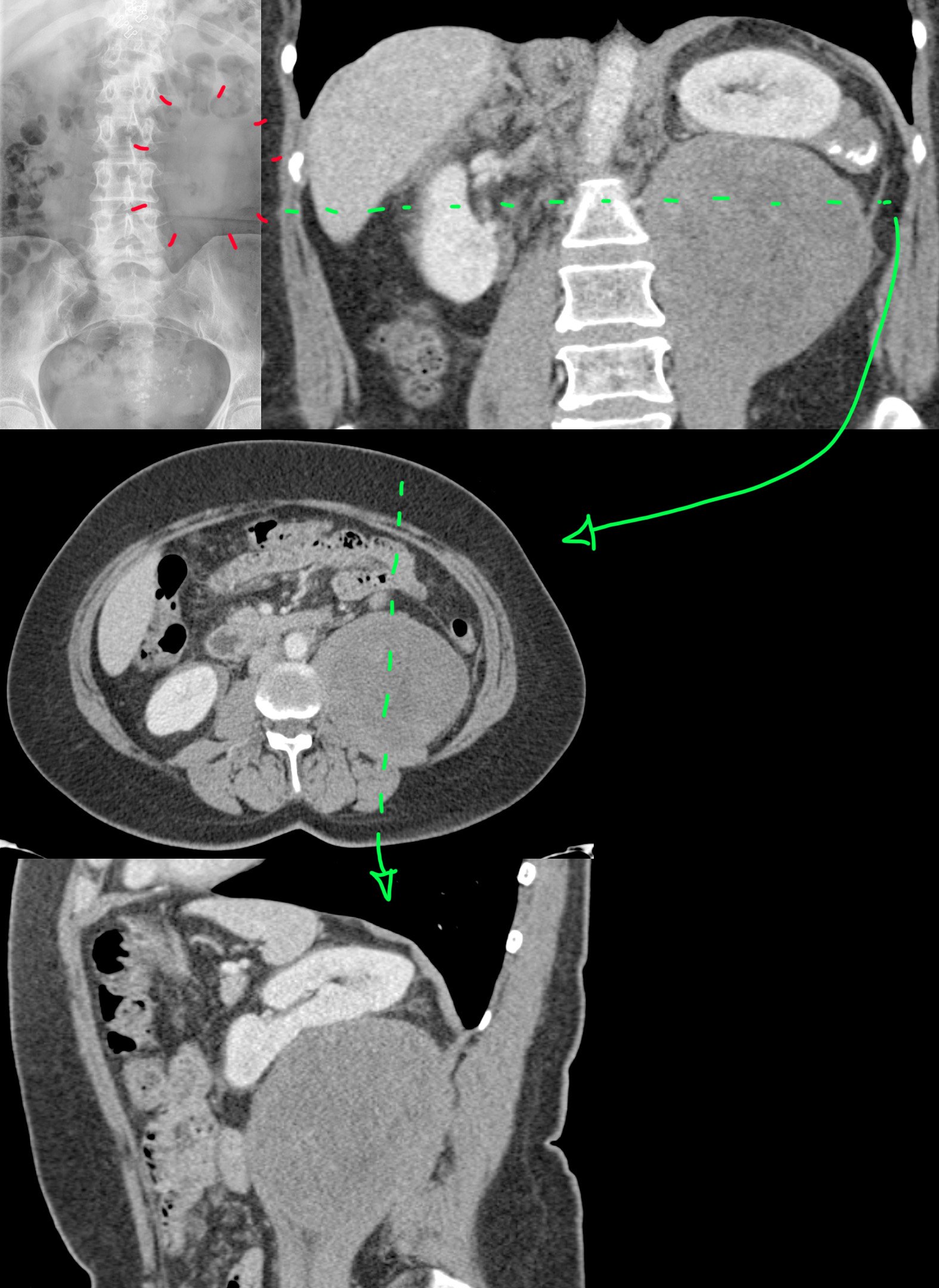
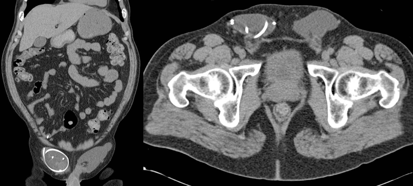
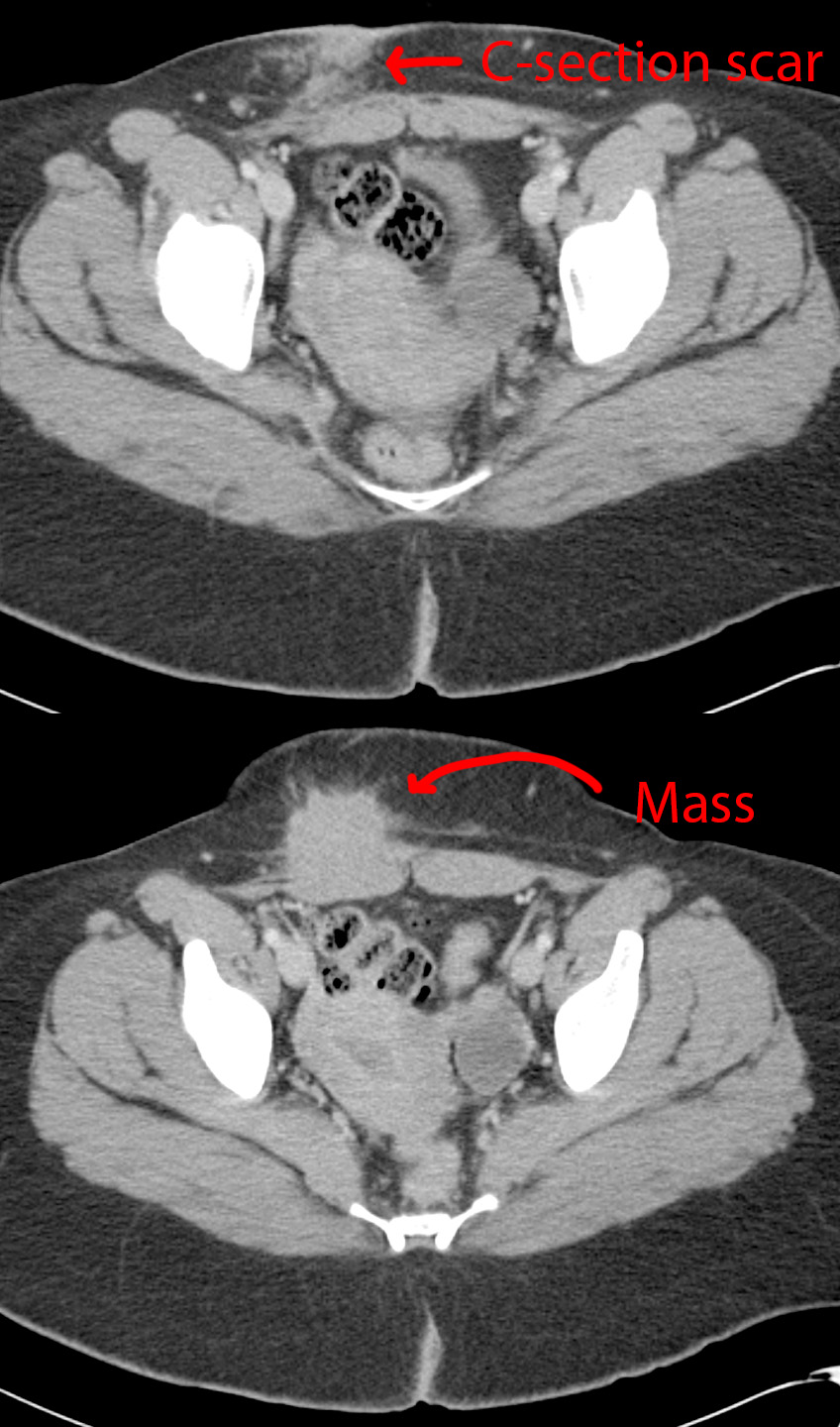
Radiology

594 readers
3 users here now

A community for all things related to medical imaging!

RULES:

-1. Please follow the Lemmy.World Server Rules.

-1A. Please be civil.

-1B. Please be respectful when discussing medical cases. While we do not wish to impose a somber tone upon this community, please remember that there are real patients behind the images.

-2. No patient-identifiable information. There is zero tolerance for breaching patient confidentiality laws. De-identified information is allowed under HIPAA, the US patient confidentiality law. Consent is not required when posting de-identified information.

-3. No requests for medical advice or second opinions. Please go to your physician/provider for assistance. Online strangers will never know your clinical history as well as your actual healthcare team, nor will the images posted here be of sufficient quality or completeness for diagnostic interpretation. Any content or discussions in this Community should be considered for educational purposes only, and their accuracy or quality with regards to standard of care cannot be guaranteed.

-4. No spam or advertising. Products or companies that are mentioned as a natural course of discussion are allowed.

-5. Please do not spread misinformation.

-6. Moderators have final say in their decisions. Please, no rules lawyering.

Posts:

Only moderators may post in this community pending Lemmy's implementation of a moderator-approval process. Until that happens, you may post general comments or questions in the stickied megathread. You may also request the moderators to post on your behalf via DM, pending time and availability.

founded 1 year ago
MODERATORS
26
1
submitted 1 year ago* (last edited 1 year ago) by Spectator@lemmy.world to c/radiology@lemmy.world


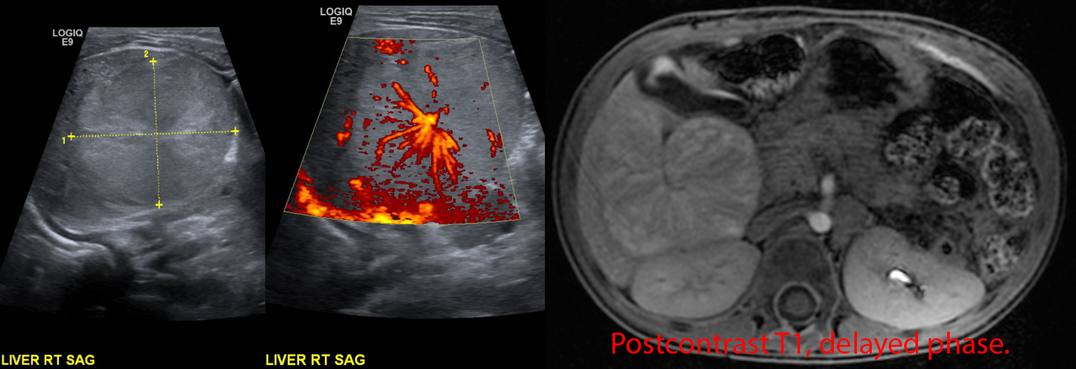
25 year old with right flank and pelvic pain for 2 days. No fevers, chills, nausea, vomiting, or other urinary symptoms however.

[Top]: Ultrasound shows right kidney with distended pelvis (hydronephrosis).

[Mid & Bottom]: Ultrasound transverse and sagittal images shows a 4.5 mm stone at the very end of the ureter (ureterovesicular junction).

Pretty straightforward case. For the nonmedical visitors, this is what we look for if your doc wants to get an US for flank pain / kidney stone suspicion. See the other case for a CT version.

27


Mom was a 36 year old with active meth abuse, homelessness, and scant prenatal care.

Prenatal ultrasound at 36 weeks shows a highly deformed fetal brain with large fluid-filled spaces that might represent cysts or hydrocephalus. The brain contours are lobulated and irregular due to absence of an overlying skull.

She was recommended for pregnancy termination but no-showed to that procedure. She then presented 3 weeks later, at 39 weeks, in labor. The baby was delivered spontaneously. The baby had absent skull above the level of the ears. The brain was large and could be seen through a membranous sac. The baby passed away one day later.

Acrania is the absence of a skull. Exencephaly is the condition where a brain (which may have otherwise been normal) is not covered by the skull in the prenatal setting. Due to the exposure of brain tissue to amniotic fluid, which is toxic to brain, exencephaly will invariably lead to anencephaly, which is the absence of all/most of the brain.

28
1
Drunk Driving (lemm.ee)
submitted 1 year ago* (last edited 1 year ago) by Kittybeer@lemm.ee to c/radiology@lemmy.world


20-something year old male. Life will never be the same for him.

29


31 year old doing some doing roof work when he fell. Seen in the ED with right ear bleeding and right-sided decreased hearing.

The normal left side is depicted in the right images (standard convention). Coronal view [top right] shows the middle ear ossicles (arrow) in the middle ear cavity, and axial view [bottom right] shows the normal "ice cream cone" appearance (arrow).

By comparison, the right middle ear ossicles are missing in the middle ear cavity [top left] (arrow with ?) and found in the external auditory canal (arrow with !!) instead. Axial view [bottom left] shows a distinct lack of the ice cream cone appearance. The middle ear and external ear canals are also filled with fluid/blood. The tympanic membrane is clearly ruptured since the middle ear ossicles are outside.

30


54 year old female with a history of uterine fibroids with persistent abdominal distention despite being told her fibroids shrunk after menopause - presumably via some pelvic ultrasound, which we didn't have.

Abdominal X-ray [top left] was possibly was called normal, which is not completely unreasonable. However, with the help of the follow-up coronal CT [top right], one can see that there is a vague area lacking any bowel gas that looks like it could be a mass. Axial [middle] and sagittal [bottom] CTs show a nonspecific, solid mass, well-circumscribed, with no calcifications, cysts, hemorrhage, or fat inside. The left kidney is pushed up by the mass, but the fat plane between the two is preserved with no evidence of renal invasion.

The patient underwent surgical resection.

Final diagnosis: Retroperitoneal dedifferentiated liposarcoma.

31


20 year old with a history of trisomy 21 and recent travel to Vietnam. Was seen there for nausea, vomiting, and abdominal pain with diagnosis of acute pancreatitis and what looked like lymphoma (but negative biopsy).

Coronal CT [left]: Big retroperitoneal lymph nodes with central cystic change/necrosis (red arrows).

Axial CT [right]: Done at the level of the green line shows a sausage-shaped pancreas compatible with ongoing versus resolving pancreatitis (red arrows).

Repeat EGD/EUS with celiac lymph node biopsy shows caseating granulomatous inflammation with positive culture and PCR for tuberculosis.

32


Not the best quality, but this is the only image I saved of this fetal MRI, with a random internet picture of what it would look like after birth.

33


This is an absolutely insane case that made the NEJM rounds a couple years ago now. I wish this was mine. I don't think there's been another case report like it since.

41 year old Colombian male with HIV-AIDS (not adherent to treatment), tapeworm infection, with several months of fever, cough, fatigue, and weight loss.

Top left: Chest x-ray shows multiple round lung masses.

Top right: Chest CT shows those round lung masses. Look like metastases.

Bottom left: The tumors taken out.

Bottom right: Pathology of the tumors.

The patient underwent empiric treatment for tapeworm and HIV with progression of masses. The CDC was contacted for help when treatment wasn't working.

The tumor cells clearly looked like cancer but were too small to be human cells. Genetic sequencing ultimately showed tapeworm DNA, with mutations and genetic rearrangements compatible with cancer.

Final diagnosis: Metastatic tapeworm cancer, likely developed because the patient had no immune system from the HIV-AIDS.

The patient passed away a few days after the diagnosis was finally made.

34


52 year old with a remote history of a rare skin cancer, dermatofibrosarcoma protuberans (DFSP) now presenting with recurrent pneumothoraces and difficulty breathing.

Top: Frontal and lateral chest radiographs shows the lungs filled by numerous cysts of varying sizes. Some cysts have layering fluid in them forming fluid levels.

Bottom: Axial CT through the mid lungs shows the numerous cysts, some with layering fluid, some with wall thickening.

Differential diagnosis for cystic lung disease: airway disease (emphysema, bronchiectasis from various causes), infectious/inflammatory (abscesses, vasculitis), cellular proliferation (cystic metastases, papillomatosis, mesenchymal cystic hamartomas, eosinophilic granulomas, lymphangioleiomyomatosis), injury (traumatic cysts, old lung infarcts).

Final diagnosis: The history of a histiocytic skin cancer suggested an extremely rare entity for cystic lung disease called pulmonary cystic fibrohistiocytic tumor.

It is sort of unclear if the lung cysts represent metastases from the fibrous histiocytic skin cancer or a separate primary neoplasm. The disease has an indolent progressive course. There is no good treatment for this as far as I am aware. These patients also get pulmonary arteriovenous malformations that might need embolization - you can see this patient does have a few embo coils on the chest radiographs at the left lung base.

35


59 year old with no past medical history, now with new dyspnea on exertion.

Top: Chest radiographs show development of cardiomegaly since 6 years prior. The pulmonary vascular markings are also more prominent than before, suggesting pulmonary congestion.

Bottom: MRI Cine FIESTA sequences in the 4- and 2-chamber views at end-diastole. There are prominent trabeculations towards the cardiac apex at both the left and right ventricles. In particular, there is an outer layer of dense myocardium and a thicker inner layer of trabeculated myocardium.

Final Diagnosis: Cardiac noncompaction.

Comparison Case: Meth-Induced Cardiomyopathy.

Cardiac noncompaction is a genetic disorder leading to failure of the myocardium to form "compact" walls. It is extremely rare. The presentation depends on how bad the noncompaction is, so can be asymptomatic to heart failure, arrhythmias, and thromboembolism.

36


20 year old with HIV-AIDS and tuberculosis with intracranial involvement (TB meningitis), not on treatment for these things.

Appropriate treatment was started, including TB medications and anti-retrovirals for HIV. 2 weeks later, the patient had decreased levels of consciousness.

Top images [A] show the brain at 3 different levels on postcontrast MRI prior to treatment. Bottom images [B] show the follow-up MRI at the time of worsening neurologic status. There is a clear progression of nodular leptomeningeal enhancement throughout the brain.

Taking the above findings into context with the time of treatment, this was felt to be consistent with immune reconstitution inflammatory syndrome, an exaggerated immune response to a co-infection (TB) when a person's immune deficiency (HIV-AIDS) is corrected. High-dose steroids were started with significant improvement. One month later, a steroid taper was attempted, but the TB-IRIS relapsed. Ultimately, it took a year of slow taper to get the patient off the steroids while continuing to treat the HIV and TB.

37


66 year old with progressive cognitive and neurological decline, becoming catatonic within 2 months.

MRI DWI [1, 2, 3] shows cortical ribboning (cortex diffusion restriction [1]), and diffusion restriction in the deep gray nuclei and insula [2, 3] in a symmetric fashion.

MRI FLAIR [4] shows abnormal bright signal at the sites of diffusion restriction.

MRI postcontrast T1 [5] and SWI [6] are normal.

Based on the above findings, there was a strong suspicion this was something bad. A lumbar puncture was performed under appropriate infection control guidelines. Results showed positive CSF results for 14-3-3, T-tau protein, and RT-QuIC. The patient had already been discharged to a nursing facility when these results came back, so the nursing facility was notified as well as the public health department. The patient was placed on comfort care.

Final diagnosis: sporadic Creutzfeldt-Jakob disease (CJD).

38


41 year old female who was stabbed through her right inframammary fold (aka under her right breast).

CT through 3 sequential phases shows a linear laceration through the right lobe of the liver. There is a small focus of contrast extravasation that enlarges over time, eventually spilling out of the liver into the peritoneal cavity. These findings are consistent with an AAST grade IV liver injury.

Oh, the knife also apparently went through the liver and diaphragm, into the chest cavity, because she also had a hemopneumothorax.

Her blood counts were trended and were stable, so she didn't need surgery for the liver laceration. She did have a chest tube placed for the pneumothorax.

39


Photo: 8 year old with a mass at the top of his head since birth.

Top left: Sagittal T1 shows a pedunculated mass protruding through the midline calvarium.

Top & bottom mid: Sagittal postcontrast T1 SPGR shows the relation of the hairpin-shaped vessel and the superior sagittal sinus to the scalp mass. There is a fenestration (hole) in the superior sagittal sinus through which the fibrous stalk courses.

Top right: Sagittal T2 shows a hairpin-shaped vessel directed towards the lesion.

Bottom right: 3D MR venogram shows the hairpin-shaped vessel pointing towards the lesion.

Atretic parietal cephalocele is a congenital abnormal herniation of the brain covering +/- brain through a midline defect of the parietal bones. It is atretic because this herniation then atrophies until only a fibrous stalk connects it to the rest of the intracranial compartment. It is associated with a persistent falcine sinus, an embryonic vertical positioning of the straight sinus.

Radiopaedia article on it.

40


A companion case to this one. Bilateral groins with fat-containing hernias. The right-sided hernia also has a small air-filled tubular structure consistent with an appendix. There are no inflammatory changes to suggest acute appendicitis, so this is an incidental finding.

A right inguinal hernia containing an appendix is called an Amyand hernia and apparently pretty rare at <1% of inguinal hernias.

41


45 year old with a ventriculoperitoneal shunt (VPS) for history of toxoplasmosis. Incidental finding - no symptoms.

CT shows bilateral groin fluid collections. The right fluid collection contains the distal loops and tip of the VPS. So, just an incidental bit of cerebrospinal fluid in the groin.

42


36 year old female with a history of C-section. Then she started feeling a growing mass in her surgical scar.

CT shows a spiculated mass along the C-section scar that invades the right inferior rectus muscle. A differential diagnosis of desmoid tumor, endometriosis, and soft tissue sarcoma was provided.

Surgical resection of the mass was performed, which turned out to be endometriosis.

43
 
 
44


Top left and right: X-rays showing a BIG right jaw mass. Whispy calcifications likely represent spiculated periosteal reaction, a very aggressive bony reaction.

Bottom left: CT in the bone/calcification window shows the periosteal reaction better.

Bottom right: MR in the T2 sequence shows the full extent of the jaw mass and how it extends far beyond the mandible's margins, both outward and inward into the floor of mouth.

While this mass is very large, in general, jaw osteosarcoma has a better prognosis than conventional osteosarcomas.

45
 
 
46


Two different patients, both aged 59, with known multiple myeloma.

47


50 year old female with history of alcohol abuse.

Baseline (6 months before): CT shows diffuse moderately low attenuation liver compatible with (alcoholic) fatty liver disease.

Patient was admitted for septic shock. CT shows a markedly enlarged liver (compare the left margin of the liver [red arrows]), markedly decreased liver attenuation, and engorged portal veins.

Subsequent CTs show the diffusely low liver evolving into a heterogeneous, splotchy, appearance and development of edema [green arrows] from decompensated liver failure. Her total bilirubin rose from baseline 0.4 to 16.4 at the 2 week mark.

48


28 year old male with a history of complex congenital heart disease presenting with altered mental status.

CT [top right image]: 4 cm centrally necrotic mass and prominent surrounding brain edema. Ddx: abscess versus tumor.

DWI [top left image]: True diffusion restriction within the central part of the mass, suggesting pus.

T2 [bottom right image]: Great example of the bright-dark-bright arrangement of abscesses on the T2 sequence (bright center, dark abscess capsule, bright surrounding edema).

Postcontrast T1 [bottom left image]: The abscess capsule appears as rim enhancement. The thin medial wall of the abscess [orange arrow] is growing every closer towards the adjacent lateral ventricle [blue arrow] and will ultimately rupture into the ventricle, causing ventriculitis, a high mortality situation.

This patient required surgical drainage of the abscess. The bacteria responsible was a Streptococcus species. Congenital heart disease is a risk factor for cerebral abscesses.